
Brachial Plexus Injuries
The Brachial Plexus is a set of nerves that give sensation and provide muscle control for the entire arm. Four cervical (neck, C5-8) and one thoracic (chest, T1) nerve roots emerge from the spinal cord and combine to form the upper trunk (C5-6), the middle trunk (C7), and the lower trunk (C8-T1). These nerve roots can be injured at birth during a difficult delivery where the baby gets stuck (shoulder dystocia), or from any injury where the head and the shoulder are forced in opposite directions. Stretch injuries can be mild (neuropraxia), moderate (axonotmesis), or severe (neurotmesis or avulsion). Mild stretch injuries, which are the most common, resolve within 2-3 months of injury with no lasting consequences. In football, these injuries are called “stingers” or “burners.” Moderate injuries typically begin to recover within 5-6 months. More severe injuries are unlikely to recover without surgery. The most important thing to know about Brachial Plexus injuries is that if surgery is required to fix the nerves, it should be performed within 3-9 months, depending on the type of injury, to get the best results.
Plexus 101: Anatomy and function
This video covers brachial plexus anatomy and function. Four cervical (neck, C5-8) and one thoracic (chest, T1) nerve roots emerge from the spinal cord and combine to form the upper trunk (C5-6), the middle trunk (C7), and the lower trunk (C8-T1).(1) Each of the trunks then breaks up into two divisions, one anterior (to the front) and one posterior (to the back). All of the posterior divisions come together to form the posterior cord. The anterior divisions of the upper and middle trunks come together to form the lateral cord, and the anterior division of the lower trunk continues on as the medial cord. These cords travel with the axillary artery (the major blood supply to the entire arm) and begin to give off branches. Unlike what has been described previously both in illustrations and in print, the posterior cord is actually posterolateral (to the back and on the side away from the body), the medial cord is posteromedial (to the back and closer to the body), and the lateral cord is directly anterior (in front of the artery).(2)
Plexus 102: plexus injuries
The mechanism and types of injuries that the brachial plexus can sustain are discussed in this video. Stretch injuries can be mild (neuropraxia), moderate (axonotmesis), or severe (neurotmesis or avulsion). Mild stretch injuries, which are the most common, resolve within 2-3 months of injury with no lasting consequences. Moderate injuries typically begin to recover within 3-5 months. More severe injuries are unlikely to recover on their own. There are important differences between brachial plexus birth injuries and later traumatic brachial plexus injuries.
Brachial plexus injuries not only affect the motor nerves, but also the sensory nerves. This included sensory nerves to the muscles, known as “muscle afferents.” Muscles contain sensory organs called spindle fibers, which allow the muscle to recognize stretch. Muscles in babies not only stretch when the nearby joint is moved, but also as the child grows. Muscles need to grow in response to the growth of the bones. The signal to grow and elongate is mediated by the spindle fibers, with intact spindle fibers required for optimal growth. Spindle fibers are not fully developed at birth, but themselves grow and elongate over the first few months of life. Injury to the nerves innervating the spindle fibers at birth results in cessation of development of the spindle fibers. Even if the spindle fibers re-innervate via spontaneous or surgically-assisted nerve recovery, the spindle fibers will not develop fully. Stunted spindle fibers do not allow the muscle to “feel” stretch effectively, stunting the drive for muscle elongation.(1) As a result, the muscles cannot keep up with the growth of the bones, leading to joint contractures.
Plexus 201: plexus examination
Examining an infant requires observation and stimulation. Neonates will not respond to commands, but much can be learned by watching them play and respond to a rattle or their other favorite toy. Gentle touches to the palm will often incite finger flexion. Stroking the back of the hand will incite finger and wrist extension. Remain calm, be patient, and keep your eye on the child, as they often tend to do what you want them to do as soon as your back is turned.
Plexus 202: diagnostic studies
We do not recommend routine use of expensive and unnecessary diagnostic studies such as MRI or electrodiagnostic studies (EMG/NCV). They are neither prognostic nor helpful for determining treatment, with some exceptions. We rely almost exclusively on clinical examination, with diagnostic testing reserved for instances where clinical examination is inconclusive. An MRI of the cervical spine should be reserved for children with complete injuries (C5-T1) who are about to undergo surgery, primarily to distinguish between avulsions and ruptures. An MRI of the brachial plexus itself is not recommended.
Starting at 2 months of age, children with limited shoulder motion should have an ultrasound of the shoulder. The ultrasound is a reliable method to evaluate the degree of glenohumeral dysplasia. Glenohumeral dysplasia can range from mild, with slight retroversion of the glenoid, to severe, known as an infantile dislocation. Glenohumeral dysplasia has been shown to develop in children as young as 2 months of age. The technique is simple to perform in an office setting and does not cause any pain to the child. Ultrasound is a reliable screening tool but does not visualize the glenoid very well. For children over 1 year of age, we recommend an MRI of the shoulder using axial images with cartilage sensitive sequences. X-rays and CT scans are not useful in children under 8 years of age because the shoulder joint is primarily cartilage in younger children.
Plexus 301: early treatment
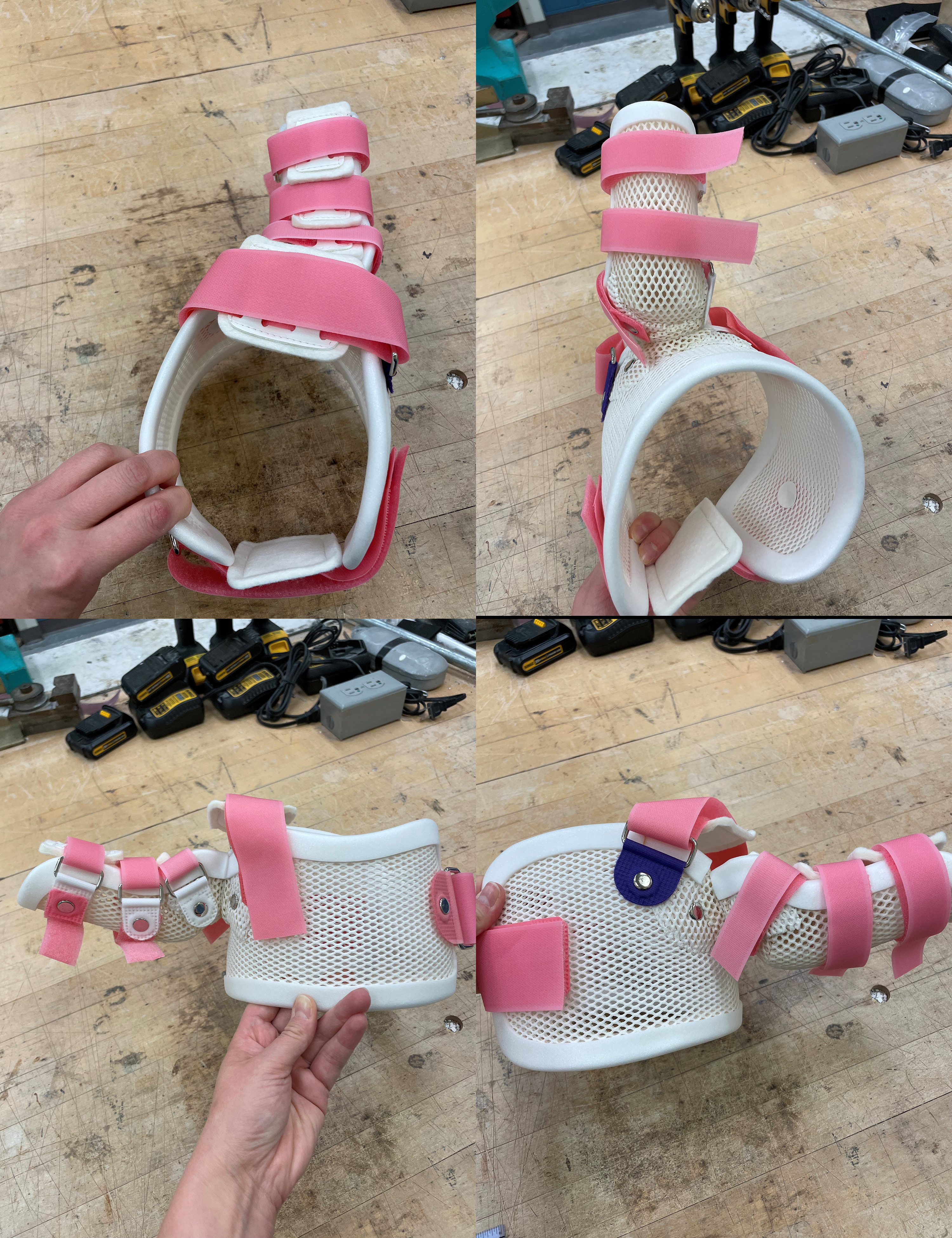
Immediately after the injury, it is appropriate to give the arm a rest by safety pinning the sleeve to the belly of the baby’s onesie. The arm may be moved for bathing and dressing without worry of causing further injury. Do not pick up your child by the injured arm during this time. Once two weeks have passed, passive range of motion exercises should begin, both at home with every diaper change and with a therapist 1-3 times a week. The main focus is to maintain shoulder external rotation both in adduction and abduction. Night time bracing may be appropriate in some children. Torticollis is often seen with plexus injuries, and may require stretching of the neck muscles as well.
Plexus 302: surgical options
Surgery for brachial plexus injuries can be divided into nerve surgery (usually performed within 9 months of injury) and salvage surgery (surgery performed usually after nerve recovery has stabilized to improve the function of the arm or prevent or mitigate deformity and contractures). Nerve surgery is done to reconnect the brain to non-working muscles by restoring working nerves to that muscle.
Plexus 303: Elbow Contracture management
Elbow flexion contractures are very common in children with BPBI. Even if elbow flexion recovers fully, the biceps and brachial muscles often cannot grow fast enough to keep up with the growing skeleton of the child, leading to tightness of these muscles. The muscles restrict elbow extension, leading to a flexion contracture. Almost all children who recover elbow flexion after 2 months of age will have some degree of an elbow flexion contracture. We recommend passive range of motion exercises throughout childhood to stretch the elbow. If these fail and the elbow contracture worsens to more than 30 degrees, we recommend a night-time static elbow extension splint. If the splint fails and the child develops a contracture beyond 45 degrees, serial casting may be considered, with a new cast each week for 4-6 weeks or until no further gains are made. Surgery should be reserved for more severe contractures that fail casting. A multitude of elbow release procedures have been proposed, but none have so far proven better than serial casting and each carries real risks to the child’s arm.(1,2,3) Botulinum toxin (botox) has also been used in conjunction with serial casting, but its role remains unclear.(4)
Plexus 304: Shoulder contracture management
Night-time bracing is used to prevent worsening of shoulder internal rotation contractures in children with <30 degrees of external rotation. Used regularly, braces can be very effective. A new one needs to be made every 6-12 months as the child grows.
If night-time bracing is insufficient or is not possible, one common procedure to minimize shoulder deformity is injection of the internal rotators of the shoulder with botulinum toxin (botox) and placement of a shoulder spica cast. There is insufficient data to know whether the cast or the botox alone are each effective, so most surgeons will use both treatments together to maximize the chances of success. Overall, botox and casting has a 15% success rate in completely resolving the shoulder tightness and glenohumeral (shoulder joint) deformity. However, there is some data to suggest that in about 2/3rds of children, the botox and casting combination does at least improve the shoulder deformity and contracture.(1,2,3)
Management of late-presenting children or children who have failed splinting, casting, and botox treatment is highly controversial. There is no “magic bullet” that can restore normal shoulder function for these children, despite the claims of some surgeons. In general, with all other factors being equal, the milder the contracture and the younger the child, the better the results with surgery.
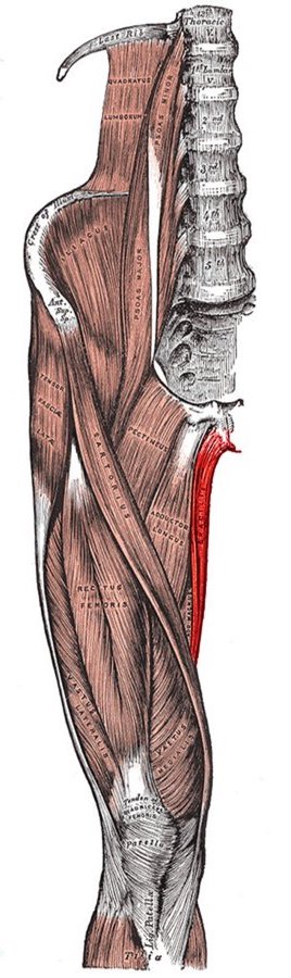
For children with fixed contractures, contractures that cannot be passively corrected, a joint release is required. Most children with brachial plexus birth injuries have internal rotation contractures, particularly when the shoulder is in adduction. Tight structures include the upper portion of the subscapularis and the anterior capsule. Although some surgeons will routinely release the pectoralis major, the latissimus dorsi, and/or the teres major, these muscles rarely contribute to the contracture and are important to preserve to minimize the risk of loss of midline function. We will only release these muscles if the child is tight in external rotation and 90 degrees of abduction, beginning with the tendinous portion of the pectoralis major. The muscular attachment of the pectoralis major to bone is preserved to maintain function and cosmesis of the anterior axillary fold. We have never had to release the latissimus dorsi or the teres major in order to get a tension-free glenohumeral joint reduction and contracture release. Releasing or transferring the latissimus dorsi and/or the teres major muscles does increase the risk of losing midline function and of developing an external rotation contracture.(4) Most surgeons in the US and internationally have therefore abandoned the classic Hoffer procedure (5) or its derivative 4-part modifications, although good results are possible with any procedure that incorporates a release in the right patient.
There are myriad options for anterior release of the shoulder (glenohumeral) joint, including arthroscopic and open techniques. The arthroscopic reduction has the advantage of leaving only two small scars which are nearly imperceptible, but it does have a potentially higher risk of nerve injury than the open techniques. The arthroscopic technique as currently performed also does not remove the coracoid. The coracoid is a bony prominence (still cartilage in young children) that serves as an attachment point for the coracohumeral ligament and the pectoralis minor, coracobrachialis, and short head of the biceps muscles. As the humeral head migrates posteriorly, the coracohumeral ligament pulls on the coracoid and deforms it into a hooked shape (6). The deformed coracoid blocks reduction of the glenohumeral joint, forcing the humeral head inferiorly and resulting in an abduction contracture. We therefore favor an anterior open approach with coracoidectomy, and have seen better results with this technique than with all others (see video).(7)
Plexus 403: brachial plexus surgical exposure
The brachial plexus can be explored at any age via a transverse supraclavicular exposure. The incision heals very nicely and is often not noticeable concealed within the natural skin creases of the neck (Langer’s lines). A superior longitudinal extension, still practiced today, is unnecessary to achieve exposure of the entire plexus, including C4, and should be avoided. The longitudinal extension leads to an unsightly scar (Figure 1) and may worsen the child’s torticollis (1). The incision is planned to extend across the posterior triangle of the neck between the anterior border of the trapezius muscle and the posterior border of the sternocleidomastoid muscle. Epinephrine solution is injected under the skin before prepping and draping to aid with hemostasis. After the incision is made with a blade, the subcutaneous tissue is dissected with a monopoly electrocautery to the level of the fascia overlying the nerve branches of the cervical plexus and the external jugular vein. The nerves of the cervical plexus should be preserved wherever possible. These nerves provide sensation to the infraclavicular skin and can form painful neuromas if cut. They are often a useful landmark which can be traced proximally to where they divide from the phrenic nerve. They may also be useful as additional nerve autograft should the sural and medial antebrachial cutaneous nerves prove insufficient. Occasionally, they can be used as a source of sensory axons to aid in plexus reconstruction. For example, the nerves can be cut distally and coapted to the anterior division of the upper trunk to provide C6 sensation (thumb and index finger) in cases where there are insufficient axonal sources from the plexus nerve roots to otherwise reconstruct the anterior division of the upper trunk. In this example the primary function of the anterior division of the upper trunk, elbow flexion, would be restored with a distal nerve transfer directly to the terminal nerve branches to the biceps or the brachialis muscles.
The external jugular vein often needs to be ligated, but occasionally may be retracted medially. The fat pad covering the plexus is then encountered and mobilized laterally. The omohyoid muscle can be found within the fat pad and serves as a marker for dissection. The upper trunk is superior to the omohyoid and the lower trunk is inferior. Once isolated, the omohyoid can be safely and effectively retracted without the need for ligation. In patients with plexus injuries, the scar typically begins deep to the fat pad, but may include the fat pad as well. Dissection should be carried out carefully with early identification of the phrenic nerve and the carotid sheath. Although the phrenic nerve runs deep to a fascial layer covering the anterior scalene muscle and deep to the carotid sheath, scar that results from a plexus injury may involve the anterior scalene muscle, disrupting the normal course of the phrenic nerve. As a result, the nerve will occasionally be found within the scarred fat pad and may even take on a transverse course for a short segment. Although respiratory dysfunction is unlikely from the loss of only one phrenic nerve, even in an infant, identification and protection of the phrenic nerve is critical to prevent a high riding diaphragm and the potential formatter respiratory complications. Infants with a one-sided phrenic nerve injury will be unable to generate suction and therefore have difficulty feeding.
The carotid sheath contains the carotid artery, the internal jugular, and the vagus nerve. All three structures are critical and should not be injured. Injury scar may pull the sheath out of its normally protected position deep to the sternocleidomastoid muscle, risking injury. A scarred fat pad likewise may become adherent to the sheath and difficult to separate. Slow and careful dissection wins the day. Branches of these major vessels, namely the supraclavicular artery and vein (traversing superficial to the divisions of the brachial plexus), the dorsal scapular artery and vein (superficial to the C7 nerve root), and the transverse cervical artery and vein (superficial to the upper trunk), should be identified and ligated. We often remove segments of these arteries to aid with exposure. The nerve roots exit the neuroforamen of the cervical spine between the anterior and middle scalene muscles. The upper trunk is made up of the smaller and more cranial C5 nerve root and the larger and more inferno-posterior C6 nerve root. The C7 nerve root can be found yet more infer-posterior to C6, continuing on as the middle trunk. Further inferior, immediately deep to the subclavian artery, are the C8 and T1 nerve roots that make up the lower trunk.
Each trunk divides into an anterior and a posterior division, with the upper trunk forming a trifurcation between its two divisions and the suprascapular nerve (2). The anterior divisions of the upper and middle trunks joint form the misnamed “lateral” cord, even thought the cord is actually directly anterior to the axillary artery. The likewise misnamed “lateral” pectoral nerve is the most anterior structure at this level, emanating from the junction of and fed by the anterior divisions of both the upper and middle trunks. The anterior division of the lower trunk continues on as the misnamed “medial” cord, which is actually poster-medial to the axillary artery. The posterior divisions of all three trunks join to form the also misnamed “posterior” cord, which is postero-lateral to the axillary artery. Typically, the posterior divisions of the upper and middle trunks will join first, followed more distally by the lower trunk. However, we have seen the posterior division of the lower trunk join the posterior division of the middle trunk before they both joined the contribution from the upper trunk. We have also seen anatomic variations where C5 and C6 never combined to make an upper trunk, and instead C5 went directly to the poster-lateral cord and C6 to the anterior cord.
Intraoperative stimulation of the nerves, cords, divisions, and trunks can help to verify the anatomic structure as well as confirm injury or suggest recovery potential. We have found that an intact nerve will convey a tetanic contraction to the downstream muscle when stimulated by a Checkpoint Stimulator (Checkpoint Surgical, Cleveland OH) at 0.5 mA and 100 microseconds pulse width. If the surgical exploration is performed within 6 months of injury, stimulation at 2 mA and 200 microseconds will induce some antigravity joint motion if the nerve is injured but likely to recover on its own. After 6 months, determination of recovery depends on a multitude of factors including patient age, distance from injury site to motor endplates, nerve composition (mixed vs. motor nerve), and time from injury.
Plexus 404: Intercostal nerve transfer
Intercostal nerves can be tedious and risky to harvest. This video demonstrates the "Leiden" technique of harvesting the intercostals without the need for stripping of the ribs. This method is both faster and safer, making it better for patients and surgeons alike. The intercostals (3 at a time ideally) can be used to power the musculocutaneous, long thoracic, triceps, suprascapular, and axillary nerves.
Plexus 405: free gracilis muscle transfer
When there are no local muscles available for functional restoration of the injured arm, one option is bringing in a muscle from somewhere else. Functional free muscle transfer is different from a muscle transplant in that the muscle come from the injured person, and not from a donor. While as parents we would all willingly give one of our own muscles to help our child, this is simply not possible because of the risk of rejection of the transplanted muscle. Patients who have undergone a transplant, whether for a new donor heart, kidney, or even hands, must take anti-rejection medications for the rest of their lives. These medications carry major risks including the development of cancer and of life threatening infections.
Fortunately, there are muscles within each of us that are redundant and therefore expendable. The gracilis muscle is one of those muscles, and perhaps the best muscle for restoring arm functions.(1,2) It is a strong muscle with a stout tendon and a single long nerve and artery that is relatively straightforward to reconnect to arteries and nerves in the arm. There is no loss of function in the leg in patients without previous injury to the leg. Typically, two functions can be achieved one gracilis muscle due to the length and power of the muscle, such as combined elbow bending and grip, elbow bending and forearm rotation, or elbow straightening and wrist extension.
Although the risks are minimal, the operation does require a team of skilled surgeons about 6-8 hours to perform. The child spends the night under sedation in the intensive care unit overnight to minimize the risk of pulmonary (lung) problems. By the next day, the child is awake and recovering from surgery. Drains are placed in the leg and removed a few days later to minimize the risk of fluid collecting in the leg.
Because the nerve to the gracilis had to be cut and reconnected to a nerve in the arm, the nerve has to grow into the muscle before it can function. This can take anywhere between 6 to 24 months, but most often between 6 and 12 months. Once the nerve has grown in, the gracilis muscle begins to work on its own. Therapy can make recovery of muscle control better and more speedy, and begins as soon as the muscle begins to move. Typically, twice weekly therapy over the course of 2 months is sufficient to restore good muscle control and strength.
Plexus 406: Shoulder tendon transfers
There are myriad procedures that have been described for improving external rotation and abduction of the shoulder.
Plexus 407: Thoracic Outlet Syndrome
Compression of the brachial plexus as it enters the thorax (chest) is called Thoracic Outlet Syndrome (TOS). The most common site of compression is between the anterior and middle scalene muscles and the 1st rib, making a triangle. Further down, the nerves can be compressed by a second triangle formed by 1st rib, coracoid, and the pectoralis major muscle.