
Arthrogryposis Education Center
Arthrogryposis 101: Introduction to amc
Arthrogryposis, also known as Arthrogryposis Multiplex Congenita (AMC) is a descriptive diagnosis for any child born with stiff joints in more than one limb. There are over 300 different conditions and syndromes that fit the Arthrogryposis definition, and all 300 fit into one of 3 subcategories: Amyoplasia, Distal Arthrogryposis (DAs), and Neuromuscular Disorders. Amyoplasia is the most common single condition, making up about a third of all diagnoses. The DAs are the next most common, with approximately 15 distinct diagnoses within this category. The neuromuscular causes are the least common and include spinomuscular atrophy and titinopathy.
The cause of Amyoplasia is unknown. What we do know is that children with this condition do not fully develop their muscles. Some muscles may be a little weak, while others may be completely absent.
Arthrogryposis 301: Elbow Release
Children with Amyoplasia are often born with elbows that do not bend very well. We recommend stretching exercises as early as possible to bend the elbow, except in cases where there is less than 30 degrees of elbow bending. Bending a nearly straight elbow is problematic because it can be difficult to tell in which direction to bend. A surgical elbow release is performed on children who cannot bend their elbow past 75 degrees at 1 year of age. The optimal time to do the surgery is after 1 year of age but before 2 years of age.(1) An elbow release can improve passive motion, but does not necessarily improve active motion. On average, children can expect to gain about 40 degrees of total motion, with an average range of motion of the elbow after surgery of 30 to 100 degrees. The risks of surgery are minimal, but triceps weakness and loss of motion can occur. Nerve injury is possible but rare.(2,3,4)
Arthrogryposis 302: Humeral rotational osteotomy
Internal rotation contractures of the shoulder are common in children with Amyoplasia. The treatment for this contracture is to cut the arm bone (humerus), rotate the arm into a better position by rotating the cut bone, and then fixing the bone in the new rotation with plates and screws.(1) Combining a humeral rotational osteotomy with an elbow release has been shown to be more risky than doing the operations separately, but may be beneficial in a small subset of children.(2) Functional improvements can be dramatic, allowing the child to see the palm of their hand and to allow both palms to face each other. This not only allows for improved bimanual tasks, but children typically report improved hand function.
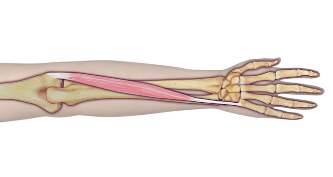
Arthrogryposis 303A: Carpal Wedge Osteotomy
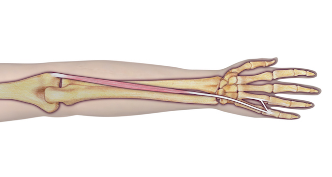
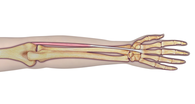
Wrist flexion contractures are very common in children with Amyoplasia. For some children, wrist flexion is functionally beneficial. Children may rely on wrist flexion for 1) reaching their mouth, 2) reaching through the legs to wipe themselves after the bathroom, 3) opening their fingers, and/or 4) weight bearing during scooting. For children without these needs, a carpal wedge osteotomy can change the position of the wrist into more extension by about 45 degrees.(1,2) The results of a carpal wedge osteotomy can vary depending primarily on the strength of the extensor carpi ulnaris (ECU) which is moved to the center of the wrist to help with wrist extension (see 303B below). The main benefit of wrist extension is that it improves grip and pinch strength. A secondary benefit is that it improves the appearance of the wrist. The results of surgery are dependent on the quality of the ECU muscle, which can vary dramatically from child to child with Amyoplasia. If the muscle is not sufficiently strong, recurrence of the flexion deformity can occur. While most children will maintain some motion at the wrist, it is common for children to lose some of their arc of motion that they had before surgery, although this is usually a loss of less than half of the motion they had before surgery. Complete stiffening of the wrist can occur but is uncommon. Surgical complications are rare but can include infection, nerve or artery injury, tendon injury, nonunion (bones don’t heal), and malunion (bones heal incorrectly). The most likely risk is recurrence. Revision of the osteotomy is not possible in the case of recurrence.
The surgery begins with a 10 cm longitudinal incision over the ulna. The flexor carpi ulnaris (FCU), flexor carpi radialis (FCR), and palmaris longus (PL) are isolated, and a strip of volar forearm fascia is removed. The ulnar and radial nerves and arteries and the median nerve are identified and protected prior to removal of any tendons or fascia. If there is no muscle on the FCU, FCR, or PL, the tendons are removed. Tendons without muscle have no ability to stretch or function, and are functionally ligaments. It is these “ligaments” that are responsible for the wrist flexion and ulnar deviation deformity in children with Amyoplasia, and these need to be removed. If there is good muscle attached to the tendons and good excursion of these tendons, then they should be preserved and fractionally lengthened as needed. Occasionally, if the ECU is of poor quality, we will transfer the FCR or even the FCU to the ECRB through the interosseous membrane if these have better associated musculature. If releasing the flexor tendons and volar fascia results in 30 degrees or more of passive wrist extension, a carpal wedge is not necessary. If passive extension is only to neutral, then a carpal wedge is recommended. In between, it is a judgment call based on the child’s needs and finger function.
The ECU is then isolated through the same incision. If not performing a carpal wedge, a separate 2 cm transverse incision is then made at the dorsum of the wrist over the ECU insertion. The dorsal sensory branches of the ulnar nerve are identified and protected. The ECU compartment sheath is opened and the ECU is released from its insertion. Be careful to identify the extensor digiti minimi (EDM), as this can be easily mistaken for the ECU. A separate 2 cm transverse incision is made over the distal aspect of the 2nd extensor compartment. The ECRB and the extensor pollicis longus (EPL) tendons are identified. The 2nd extensor compartment sheath is opened. If the ECRB has no excursion, the tendon is divided as proximally as possible. The ECU is pulled into the proximal longitudinal incision and redirected via a subcutaneous tunnel to the ECRB tendon. If a carpal wedge is to be performed, it is performed now. If not, the ECU is transferred to the ECRB via a single Pulvertaft weave and multiple side to side Krackow sutures with the wrist tensioned in maximal extension.
The wounds are closed and a sterile dressing is applied. The wrist is then casted in maximal extension in a short arm cast for 4-6 weeks. In children younger than 3 years old, a long arm cast is used. Therapy starts as soon as the cast is removed, with an intensive 3 day session followed by a home program and biweekly therapy visits.
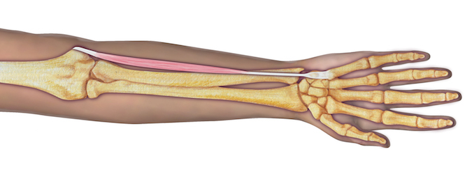
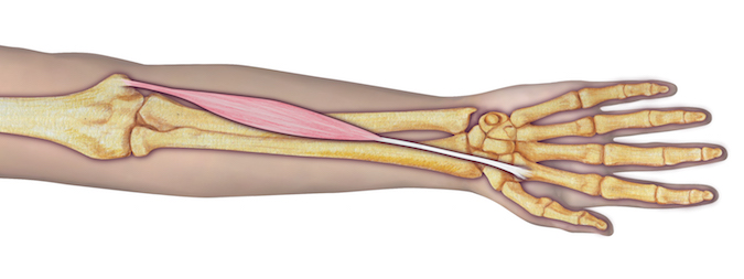
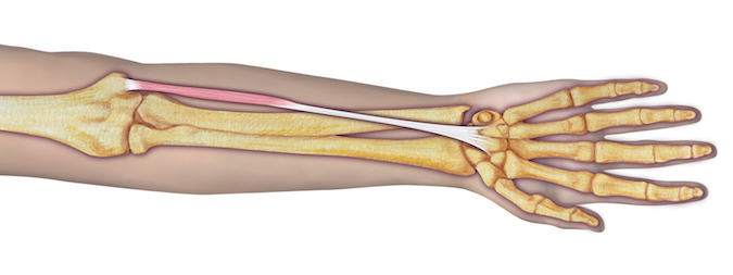
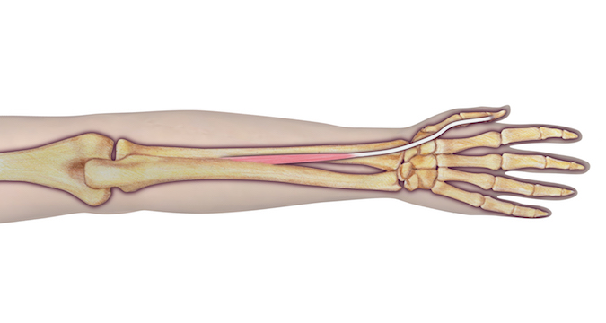
Arthrogryposis 303B: ECU to ECRB tendon transfer
For children with limited or no active wrist extension, the extensor carpi ulnaris muscle (ECU) can be moved from its position on the ulnar side of the wrist and sewn into the extensor carpi radials brevis (ECRB) at the center of the wrist. The change in position allows the ECU to work like the ECRB to extend the wrist. Children with good passive motion to about 30 degrees of extension are great candidates for this tendon transfer, provided that there are no contraindications to placing the wrist in more extension (see 303A above). These contraindications include poor finger extension, obligate wrist flexion for wiping and eating, and weight bearing on the back of the wrist for scooting. If there is inadequate passive wrist extension, the ECU to ECRB transfer needs to be performed along with a carpal wedge osteotomy (see 303A above). The main benefit of wrist extension is that it improves grip and pinch strength. A secondary benefit is that it improves the appearance of the wrist. The results of surgery are dependent on the quality of the ECU muscle, which can vary dramatically from child to child with Amyoplasia. If the muscle is not sufficiently strong, recurrence of the flexion deformity can occur. It is also possible for the wrist to get stuck in extension, although this is rare. Surgical complications are rare but can include infection and nerve or artery injury.
The wounds are closed and a sterile dressing is applied. The wrist is then casted in maximal extension in a short arm cast for 3 weeks. In children younger than 3 years old, a long arm cast is used. Therapy starts as soon as the cast is removed, with an intensive 3 day session followed by a home program and biweekly therapy visits.
Arthrogryposis 304: Thumb reorientation Osteotomy and stiletto flap
Children with Amyoplasia and those with a Distal Arthrogryposis typically have different thumb contractures.(1) For children with Amyoplasia, the typical thumb position is to bend at the base of the thumb. Children with Distal Arthrogryposes tend to have extension at the base of the thumb and flexion at the middle thumb joint (metacarpophalangeal joint). For children with the typical Amyoplasia thumb posture of flexion at the base (carpometacarpal joint) of the thumb, an extension osteotomy can bring the thumb out of the palm, improving pinch and grasp. Since most children with Amyoplasia also have a supination rotational deformity of the thumb, where the thumb is more in the plane of the fingers, a pronation rotational correction can be added to the extension osteotomy. This rotational correction allows the pulp of the thumb to contact the pulp of the index finger, rather that the side or nail of the thumb contacting the index finger. The index-based stiletto flap allows for loosening of the skin in cases where the skin is too tight to allow adequate extension of the thumb. Surgical complications are rare but can include infection, nerve or artery injury, nonunion (bones don’t heal), and malunion (bones heal incorrectly). The most likely risk is recurrence, which is expected in children less than 7 years of age and common below the age of 10. Revision of the osteotomy is possible in the case of recurrence.
Arthrogryposis 305: MP Chondrodesis and stiletto flap
Children with distal arthrogryposis often are born with a clasped thumb, where the metacarpophalangeal (MP) joint of the thumb is stuck in a bent position (flexion contracture). If the thumb remains in this position, the child will have difficulty grasping objects, and pinch will not be possible. Treatment begins shortly after the child is born, with passive stretching. Once the child is old enough to fit into a splint, usually between 3 and 6 months of age, a custom splint is used during naps and nights to stretch the thumb. If the thumb flexion contracture does not resolve with splinting and stretching after 2 years, the child can be considered for a metacarpophalangeal chondrodesis and stiletto flap.
The stiletto flap is a modified Z-plasty that transfers the extra skin from the radial side of the index finger into the base of the thumb MP joint and the 1st web space. Full thickness flaps are elevated to minimize the risk of losing the narrow flaps. The dorsal sensory branch of the radial digital nerve of the index finger and the princeps pollicis artery are encountered and protected. To augment thumb abduction and extension, the fascia overlying the 1st dorsal interosseous muscle and the adductor pollicis is incised.
A dorsal incision at the MP joint is made and carried down between the extensor pollicis brevis (EPB) and extensor pollicis longus (EPL) tendons. The joint is opened sharply and the collateral ligaments are divided. Enough of the metacarpal head is removed to allow for tension-free extension of the thumb. The articular (joint) cartilage at the base of the proximal phalanx is then removed sharply, with care to preserve the growth plate (physis). Fixation is achieved with a single or occasionally 2 0.045” Kirschner wires (K-wire).
A thumb spica cast is applied and kept on for 4 weeks, when the pin (K-wire) is removed in the office. The cast is then removed and a splint is worn for a further 2 weeks full time except for bathing. The splint is worn at night and naps for an additional 6 weeks.
Arthrogryposis 306: Bipolar latissimus muscle Transfer
When available, the bipolar latissimus transfer is by far the best way to give a child with Amyoplasia the ability to bend their elbow actively (with their own power). Other options for providing active elbow flexion include, in order of preference: the free functional gracilis muscle transfer, the partial pectoralis muscle transfer, the partial triceps transfer, and the Steindler flexorplasty. Although transfer of the entire triceps continues to be performed, even in the US, the operation has been shown to have poor outcomes and should not be attempted.
Arthrogryposis 307: Partial Pectoralis muscle Transfer
The partial pectoralis transfer is another option for providing active elbow flexion in children with Amyoplasia, although not nearly as reliable, successful, or as strong as a latissimus transfer.

MRI of thigh showing fatty replacement of gracilis muscle. This muscle is not viable for transfer.
Arthrogryposis 401: Functional Free Muscle transfer (gracilis transfer)
When there are no local options for gaining active elbow flexion, a muscle from the leg can be brought up to the arm using microsurgical techniques. In order to do this, three things must be present.
1) There needs to be a good gracilis muscle in the leg. This is not possible to test clinically, and requires an MRI of the thigh.
2) There needs to be a good nerve in the arm that can be sacrificed and transferred to the gracilis muscle. This means that one muscle in the arm will have to lose its nerve supply, and therefore will lose its function. This may be intercostal nerves or the spinal accessory nerve.
3) There must be good passive elbow range of motion of at least 30-100 degrees.
Additionally, use of the gracilis muscle can weaken leg adduction, affecting gait if it is harvested. In patients with marginal musculature in the leg, we advocate temporarily paralyzing the gracilis muscle with 0.25% Bupivacaine anesthetic to see if removal of the gracilis would negatively affect gait.
Very few patients will meet the requirements to undergo this procedure, but for patients who are good candidates the results can be very good. Published series have demonstrated favorable outcomes.