
Trauma Learning Center
Trauma 101: reading and interpreting x-rays
There are a multitude of ways to describe X-rays and X-ray findings. Displacement refers to whether or not, and to what degree, two fracture fragments have separated from each other. If there is a fracture line but the bone otherwise looks normal, with the bone fragments still in their native position, a fracture is said to be non-displaced. If the fragments have moved away from each other, they are displaced. Typically, displacement is described as a percentage, where if the fracture fragments have shifted 50% of the width of the bone, the fracture is said to be 50% displaced. If there is no contact between the two ends of the fracture, the fracture is said to be completely or 100% displaced.

Trauma 102: Basics of fracture healing
Fractures require two things to heal: biology and stability. Fortunately, most children have biology to spare, with a robust healing response when a bone is broken (broken = fractured). Children have a thick bone-forming covering called periosteum that in some cases can completely grow a new bone. As long as either the fractured bone fragments are touching or the periosteum from the fragments is connecting the fragments, the fracture will most likely heal if immobilized or fixed appropriately. Bone is one of the few tissues in the body to heal without scar.
When a bone is broken, the inside of the bone (cancellous bone) is exposed, leading to some internal bleeding. This blood from the bone is filled with bone forming cells called osteoblasts. The blood clots into a gelatin-like consistency. If left undisturbed with adequate immobilization, over the next 2 weeks the osteoblasts inside the blood clot (hematoma) begin to form a cartilage matrix known as “soft callus.” Over the subsequent 2-4 weeks, the soft-callus converts to a hard callus as calcium and phosphate are deposited into the cartilage (ossification). By 4-6 weeks after the fracture occurred, the hard callus has become rigid enough to be nearly as strong as the original bone. This entire process is known as secondary bone healing, because the bone heals through an intermediary cartilage phase.
Primary bone healing, where bone heals directly to bone, is only possible when a fracture is non displaced or when it has been surgically fixed with rigid fixation such as a bone plate and screws. For primary bone healing to occur, the bone ends must be less than 1mm apart from each other. In primary bone healing, special multinucleated bone eating cells called osteoclasts walk across the fracture, coring out a microscopic hole (cutting cone) across the two bone fragments. Just behind, osteoblasts follow the osteoclasts and line the walls of the cutting cone, where they begin to form new bone. Over the course of 4-6 weeks, these holes span the entire fracture and fill with bone, making the fracture literally disappear.
Good biology without adequate stability leads to a hypertrophic nonunion. Stability without favorable biology leads to an oligotrophic or atrophic nonunion. In children, where the epiphyses (ends of the bones) rely on few nutrient vessels (feeding arteries) for their blood supply, avascular necrosis (death of the bone) can occur, leading both to a nonunion and absorption of the bone. Avascular necrosis can be a devastating complication, and is most commonly due to either the injury alone or the dissection used in the attempt to fix the fracture.

Nerve injuries
Injuries to the major nerves of the arm, the radial, median, and ulnar nerves, can be divided into high and low injuries depending on the location of the injury relative to the elbow. A high radial nerve injury result in loss of wrist, finger, thumb, and possibly elbow extension. A high and low median nerve injury may look similar to their low injury counterparts, but the prognoses of these injuries are very different. While recovery of high radial nerve injuries is possible with repair of the nerve injury, recovery of function after high median, and particularly ulnar nerve injuries is less likely. This has to do with the distance between the injury site and the point at which the nerve enters and connects to its target muscle. Nerve recovery advances from the point of injury down the arm at about an inch a month, and must reach the muscle before the muscle dies at 18-24 months. Muscles in the hand are therefore harder to recover than muscles in the upper arm.
High Radial Nerve Injuries: These injuries present with a loss of wrist and finger/thumb extension. Triceps weakness may also result if the injury is near the axilla (armpit). Restoration of finger extension can be via tendon or nerve transfers. Our preferred tendon transfer for restoration of finger extension is the FCR to EDC transfer. The FCR is a better donor than the FCU because it has greater excursion and can be transferred through smaller incisions with less dissection and tissue damage.
High Median Nerve Injuries: The median nerve provides wrist flexion, pronation, finger flexion, thumb flexion, and thenar (thumb) muscle function. Injury to the median nerve above the elbow leads to loss of forearm pronation, loss of index, long finger, and thumb flexion, and loss of thumb abduction. Wrist flexion and overall finger flexion will be weaker.
Low Median Nerve Injuries: Restoration of thumb abduction when nerve recovery has failed is possible using tendon transfers. The most common is the ring FDS oppositionplasty. If this is weak or unavailable, EIP, EDQ, Huber or Camitz transfers are good options. ECU oppositionplasty is less reliable.

Oblique osteotomy for varus deformity of the elbow
Trauma: Distal Humeral varus and valgus deformities
Varus and valgus deformities of the distal humerus can result from a variety of injuries such as supracondylar humerus fractures, lateral condyle fractures, and medial condyle fractures. Most children tolerate up to 15 degrees of deformity, but for some children even mild deformities can cause loss of function or body image issues. Late complications can included elbow pain, arthritis, and instability, as well as ulnar nerve dysfunction. Fixing the deformity can often be accomplished by a single osteotomy in the distal humerus and has a very high success rate with low risk. There are many options for osteotomies and for fixation types. We favor a simple oblique closing wedge osteotomy with plate fixation for older children and pin fixation for younger children.
Trauma 301: Phalangeal Neck Fractures (strauch pinning)
Phalangeal neck fractures can be difficult to treat. We have found that the Strauch Technique is the simplest and provides for the best outcomes.(1)
Trauma 302: Supracondylar humerus fractures
Typically seen in young children below the age of 7, supracondylar humerus fractures result from a fall onto the arm from a height or at speed. Most fractures are extension type, bending backwards. The Gartner classification is used to describe the fracture based on the severity of the injury. A Gartner 1 is not angulated nor displaced, and can be treated with immobilization alone for 3 weeks. Weekly radiographs are recommended to monitor for fracture displacement in the splint or cast. A Gartner 2 is angulated, but not displaced. If there is posterior displacement with intact posterior periosteum, the fracture is classified as a Gartner 3. Completely unstable fractures with no intact periosteum are considered Gartner type 4 under the new modified classification.
Trauma 303: medial epicondyle fractures
Medial epicondyle fractures occur in children during throwing, with direct trauma, or during a fall on the outstretched arm. They are occasionally accompanied by a full elbow dislocation, a radial neck fracture, and/or a coronoid fracture. Historically, treatment indications have been based on the amount of fracture displacement, with an arbitrary 5mm of displacement designated as an indication for open reduction of the fracture. However, the amount of displacement is both difficult to determine on radiographs and is irrelevant to the need for fracture fixation. Fixation of displaced fractures should be based on whether or not the medial collateral ligament (MCL) is avulsed with the fracture fragment. If only the flexor pronator origin remains attached to the medial epicondyle fragment, but the ligament remains attached to the distal humerus, there is no need for fixation.
Nondisplaced fractures can be treated in a cast with weekly followup until union. Displaced fractures should be taken to the operating room for an examination under anesthesia if possible. An examination in the office is nearly always inconclusive because of patient guarding. Advance imaging such as CT and MRI are likewise not useful because neither are reliable methods of assessing ligamentous integrity. We bring the patient to the operating room. If the joint is unstable to vagus load, then the fracture should be fixed regardless of fragment size or location. If the joint is stable, then it is a muscle injury and only requires immobilization for 4 weeks.
trauma 304: Lateral Condyle fractures
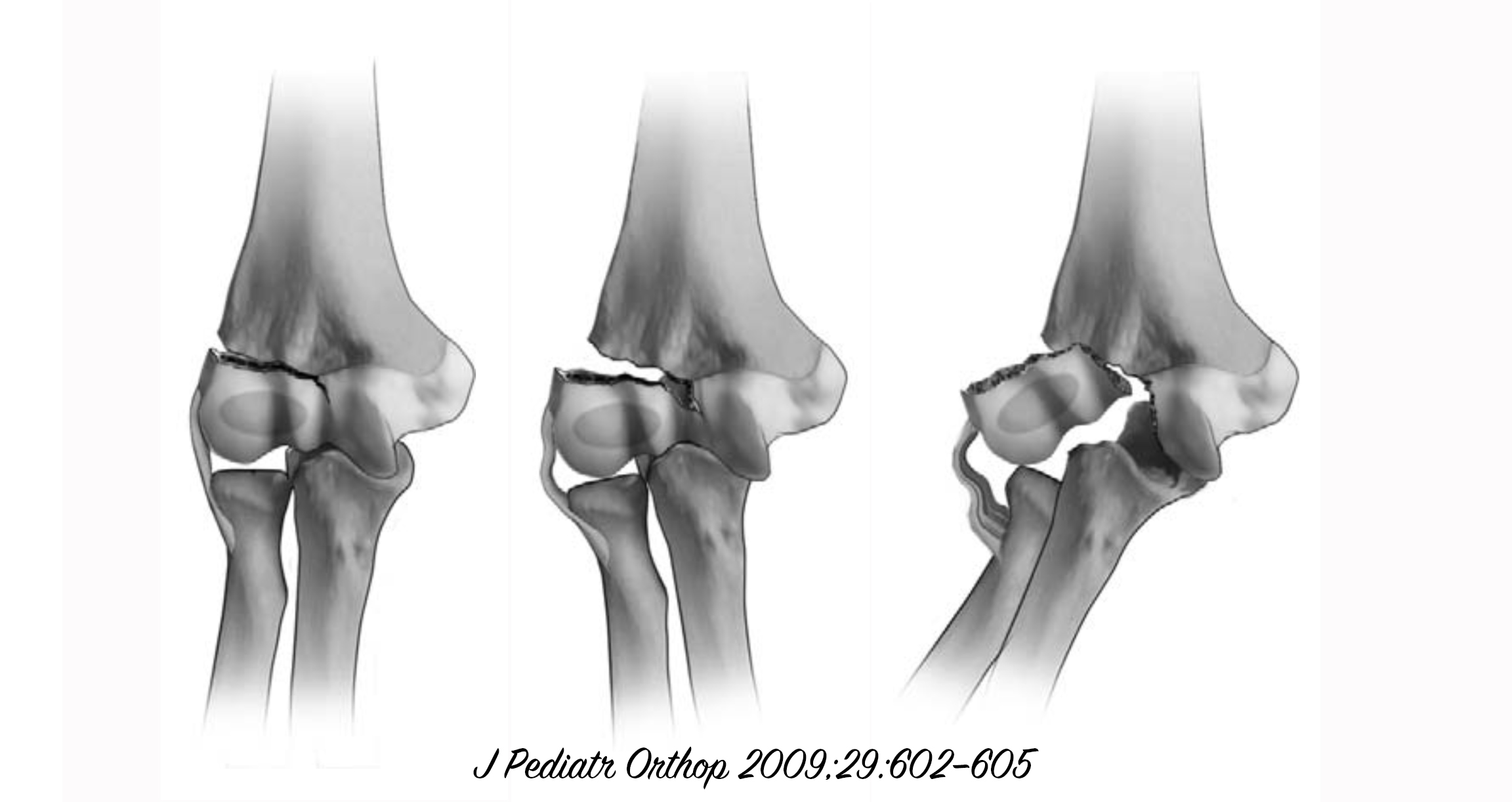
Lateral condyle fractures are the second most common pediatric elbow fracture, making up 15-20% of all distal humerus fractures in children. The average age of children who fracture their lateral condyle is 6, and the injury is often from a high energy mechanism such as a fall from a height, a bicycle accident, or a Motor vehicle collision. Because the fracture is often not visible on the AP or the lateral radiographs, lateral condyle fractures are often missed. An internal rotation oblique film is the optimal projection. The fracture is often mistaken for the lateral epicondyle, which does not ossify until after 11 years of age. Contralateral films will show the lack of an ossification center on the uninjured side and therefore simplify the diagnosis. Because the distal humerus in a 6 year old is mostly cartilage, CT scans are rarely helpful and should be avoided. An MRI is preferred if there is any doubt after internal oblique and contralateral films are taken.
There have been multiple classification schemes for lateral condyle fractures, beginning with Milch. We prefer the Weiss classification (1) for its simplicity and practicality. Type I fractures are minimally displaced (<2mm), and have an intact articular surface. Type 2 fractures are slightly more displaced, between 2 and 4 mm, but also with an intact articular surface. Type 3 fractures are more than 4 mm displaced, with a complete fracture affecting the articular surface. Type 1 fractures can be treated in a cast for 6 weeks and heal reliably if they do not displace any further. We advocate for weekly serial radiographs for the first 4 weeks and again at 6 weeks to assure that there is no further displacement. Type 2 fractures are more problematic, and are treated with an intra-operative arthrogram to confirm the classification, followed by a closed reduction and percutaneous pinning. Again, 6 weeks in a cast is recommended before pulling the pins in the office. If the fracture is complete, a type 3, then an open reduction and fixation is required to realign the joint surface anatomically. Pins are used in younger patients, but screws may be used for older patients nearing or at skeletal maturity. The pins are removed after union occurs, typically at 6 weeks.
Failure to appropriately treat these injuries can lead to a cubits valgus deformity of the elbow and tardy ulnar nerve palsy. Even with appropriate management, complications can occur. These include cubits varus and valgus deformities, with varus occurring more commonly. Avascular necrosis can also occur resulting from the initial trauma or the surgical approach. Care should be taken to preserve all soft-tissue attachments to the lateral condyle during open reduction and fixation.